Left Hip Muscles Anatomy - Hip Muscles The Definitive Guide Biology Dictionary / Included within the chart are gorgeous illustrations of the pelvic diaphragm, sphincter muscles, gluteus maximus.
Left Hip Muscles Anatomy - Hip Muscles The Definitive Guide Biology Dictionary / Included within the chart are gorgeous illustrations of the pelvic diaphragm, sphincter muscles, gluteus maximus.. It is a flat, triangular muscle on the anterior wall of the pelvis. In order to isolate the abdominals, you need to minimize the involvement of the hip flexors and maximize the contraction of the abdominals. The hip muscles encompass many muscles of the hip and thigh whose main function is to act on the thigh at the hip joint and stabilize the pelvis. In human anatomy, the muscles of the hip joint are those muscles that cause movement in the hip. Most modern anatomists define 17 of these muscles, although some additional muscles may sometimes be considered.
Included within the chart are gorgeous illustrations of the pelvic diaphragm, sphincter muscles, gluteus maximus. Rectus femoris muscle, one of the quadriceps muscles on the front of your thigh. Major lower body muscle groups include leg and hip muscles, largest muscle groups in your body. In human anatomy, the muscles of the hip joint are those muscles that cause movement in the hip. Pelvis and acetabulum, with muscle attachment sites.
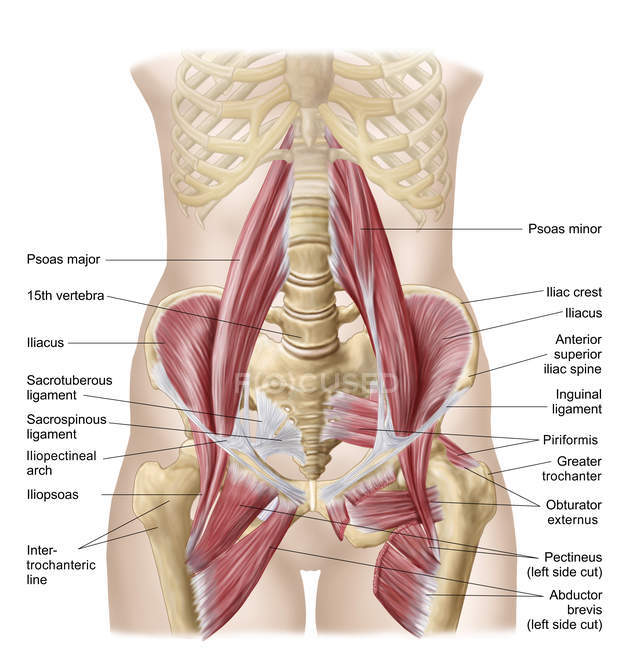
Muscles Of The Hip Wikipedia from upload.wikimedia.org In human anatomy, the muscles of the hip joint are those muscles that cause movement in the hip. The main functions of the neck muscles are to permit movements of the neck or head and to provide structural support of the head. The muscles of the pelvis, hip and buttock anatomical chart shows how each muscle in this area of the body works with the others, and the various minor systems within the major ones. Rectus femoris muscle, one of the quadriceps muscles on the front of your thigh. Microscopic anatomy of skeletal muscle. The psoas major muscle (usually shortened to just the psoas muscle) is one of the muscles of the posterior abdominal wall and lies not in the retroperitoneum but posterior to it, in the iliopsoas compartment. If you know all the hip flexor names and bones they attach to, that's an awesome accomplishment! Leave a reply cancel reply.
In clinical anatomy the thigh muscles are divided into three groups:
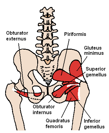
It is a flat, triangular muscle on the anterior wall of the pelvis. Muscle movements, types, and names. The muscles of the pelvis, hip and buttock anatomical chart shows how each muscle in this area of the body works with the others, and the various minor systems within the major ones. Attached to the bones of the skeletal system are about 700 named. Learn about hip muscles human anatomy with free interactive flashcards. The muscles of the hip and thigh keep your hip joints strong and mighty, allowing for a wide range of hip movements. The hip joint is the articulation of the pelvis with the femur, which connects the axial skeleton with the lower extremity. In order to isolate the abdominals, you need to minimize the involvement of the hip flexors and maximize the contraction of the abdominals. Anterior muscles extend your legs and flex your thighs. Trunk muscles, 289 muscles of the thorax, 289 muscles of the abdominal wall, 289. The cavity of the acetabulum the external obturator muscle is short external rotator muscle of hip joint. Meanwhile, labral sulcus and absent labrum are normal variations in the labrum (ring of cartilage). There are a lot of muscles of the hip and thigh.
Knee assessment and hip mechanics learn how hip and pelvis mechanics can influence the knee powered by physiopedia start course. The different anatomical areas of the gluteal region: 1 hip anatomy, function and common problems. The muscular system is responsible for the movement of the human body. The muscles of the neck can be divided into groups according to their location.
Anatomy Of Iliopsoa With Dorsal Hip Muscles Physiology Iliopsoas Stock Photo 174715594 from st.focusedcollection.com One example of an ab exercise that actually focuses. In order to isolate the abdominals, you need to minimize the involvement of the hip flexors and maximize the contraction of the abdominals. Anatomy of the muscular system. In clinical anatomy the thigh muscles are divided into three groups: Each muscle below has the bones in bold for intermediate learners and the specific bony landmarks for advanced learners. Meanwhile, labral sulcus and absent labrum are normal variations in the labrum (ring of cartilage). Your email address will not be published. The muscles of the pelvis, hip and buttock anatomical chart shows how each muscle in this area of the body works with the others, and the various minor systems within the major ones.
Your email address will not be published.
These muscles constitute the anatomical classification known as the medial compartment of the thigh. Each muscle below has the bones in bold for intermediate learners and the specific bony landmarks for advanced learners. In order to isolate the abdominals, you need to minimize the involvement of the hip flexors and maximize the contraction of the abdominals. I pulled some muscles on left hip hiking. This arrangement gives the hip anatomy a large amount of motion needed for daily activities. Most modern anatomists define 17 of these muscles, although some additional. If left unstretched, shortened hip flexors affect the position of the pelvis, which in turn affects the position and movement of the lower back. Rectus femoris muscle, one of the quadriceps muscles on the front of your thigh. Learn their anatomy efficiently and easily using kenhub's muscle anatomy and reference charts! The cavity of the acetabulum the external obturator muscle is short external rotator muscle of hip joint. Anatomy of the muscular system. The hip's essential muscles are the sartorius, rectus femoris, gluteus minimus and medius, iliopsoas, adductors, and hamstrings. In human anatomy, the muscles of the hip joint are those muscles that cause movement in the hip.
Your email address will not be published. Advanced hip flexor muscle anatomy. The hip's essential muscles are the sartorius, rectus femoris, gluteus minimus and medius, iliopsoas, adductors, and hamstrings. It originates at the anterior inferior iliac spine and just above the acetabulum of the hip bone. In order to isolate the abdominals, you need to minimize the involvement of the hip flexors and maximize the contraction of the abdominals.
Hip Labral Tear Symptoms Causes Treatments from www.clevelandclinic.org Knee assessment and hip mechanics learn how hip and pelvis mechanics can influence the knee powered by physiopedia start course. for detailed anatomy of pelvic bones, read anatomy of hip bone. Pelvis and acetabulum, with muscle attachment sites. If left unstretched, shortened hip flexors affect the position of the pelvis, which in turn affects the position and movement of the lower back. Most modern anatomists define 17 of these muscles, although some additional. It is a flat, triangular muscle on the anterior wall of the pelvis. One example of an ab exercise that actually focuses. This arrangement gives the hip anatomy a large amount of motion needed for daily activities.
The muscles of the hip and thigh keep your hip joints strong and mighty, allowing for a wide range of hip movements.
In human anatomy, the muscles of the hip joint are those muscles that cause movement in the hip. This anatomical atlas was especially designed for a specific public (radiologists, surgeons, rheumatologists and physicians specializing in musculoskeletal imaging). Advanced hip flexor muscle anatomy. Anatomy 3d atlas allows you to study human anatomy in an easy and interactive way. These muscles constitute the anatomical classification known as the medial compartment of the thigh. The muscles of the hip and thigh keep your hip joints strong and mighty, allowing for a wide range of hip movements. Learn about hip muscles human anatomy with free interactive flashcards. Several muscles cross the front of the hip and create hip flexion, pulling the thigh and trunk toward each other, but probably the most important is the iliopsoas. 1 hip anatomy, function and common problems. Included within the chart are gorgeous illustrations of the pelvic diaphragm, sphincter muscles, gluteus maximus. Anatomical terms allow us to describe the body and body motions more precisely. for detailed anatomy of pelvic bones, read anatomy of hip bone. Pelvis and acetabulum, with muscle attachment sites.


0 Komentar